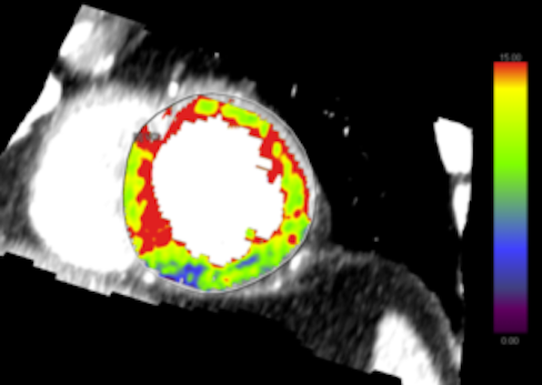
Researchers at Emory University School of Medicine recently performed a cardiac imaging procedure that was the first of its kind completed in Georgia.
The procedure, first performed on October 21, is called dynamic myocardial CT (computed tomography) perfusion imaging or cardiac CTP imaging for short. Cardiac CTP imaging is performed in conjunction with CT angiography, an X-ray scan of the heart.
“The added value is that we can provide function and anatomical assessment in one session using a single modality,” says Carlo De Cecco, MD, PhD, professor of radiology and imaging sciences at Emory University School of Medicine and director of the Translational Lab for Cardiothoracic Imaging and Artificial Intelligence.
“That means we can see both where the coronary arteries are blocked and how much – it’s kind of a one-stop shop approach,” he adds.
More conventional CT angiography provides information about partial or complete blockage (stenosis) and which arteries are involved, which aids in planning future treatment and therapy. CT angiography is currently used for the evaluation of stenosis severity and is accurate in excluding disease, but often overestimates the severity of stenosis.
Studies have shown that functional information is a better predictor of disease severity, guiding decision-making about invasive interventions such as stenting or bypass surgery. Previously procedures such as FFR (fractional flow reserve) measurement, performed during cardiac catheterization, were necessary to provide accurate functional information.
Emory researchers are conducting a study that is comparing cardiac CTP imaging to nuclear stress testing, also known as PET (positron emission tomography) myocardial perfusion imaging. The goal is to include 30 patients, De Cecco says. Valeria Moncayo, MD, assistant professor of radiology and imaging sciences, is also involved with the study, which is funded by Siemens and uses the company’s SOMATOM Force scanner.
“We are including patients who are referred for a clinically indicated myocardial PET perfusion scan,” De Cecco says. “The primary indications are cardiac-related complaints and abnormal results in initial testing, such that functional testing is required to assess for myocardial ischemia or infarction.”
In cardiac CTP imaging, a catheter is not introduced into the body, but vasodilator drugs such as adenosine/Regadenoson and iodine contrast agents are used. Cardiac CTP imaging is similar to PET-based myocardial perfusion imaging, except that no radioactive probes are introduced into the patient’s body. PET-based myocardial perfusion imaging is currently the gold standard for quantitatively assessing cardiac blood flow, but it does not allow assessment of the coronary arteries.
The actual CTP imaging exam lasts about 30 seconds. In combination with CT angiography, it can be performed in 30-40 minutes total, reducing radiation dose compared to PET-based procedures.

