In addition to world-class research, Emory scientists are producing images that could easily find homes in an art gallery or museum exhibition.
In preparation for the Postdoctoral Research Symposium this Thursday, the Office of Postdoctoral Education and the Integrated Cellular Imaging Core held a Best Image Competition. All ten of the entries can be found here. The top three, chosen by the contest organizers, were voted on by the Emory research community.
The 2017 top three winners are:
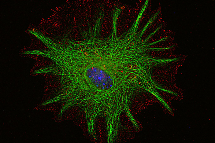
- First place: Ariel Diaz, PhD, from Manuel Yepes lab, Department of Neurology, Center for Neurodegenerative Disease
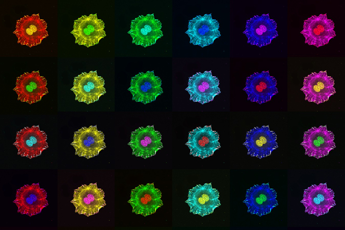
Mouse cortical astrocyte stained with anti-glial fibrillary acidic protein (green), anti-Ezrin (red) and Hoechst blue DNA stain. Second place: Alejandra Valdivia, PhD, San Martin lab, Department of Medicine, Division of Cardiology
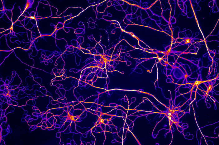
“Rainbow”. Mouse embryonic fibroblast cells were stained to visualize stress fibers (long filaments), focal adhesions (peripheral dots) and cell nucleus (double spheres at the center of the cell). Images were pseudo colored and put together in a montage to represent that beauty comes in different shades.Third place: Sandii Constable, PhD, Caspary lab, Department of Human Genetics
Cultured cortical neurons, stained for acetylated tubulin. Color depicts intensity of staining: high intensity is yellow or white, while low intensity is purple.
Winners receive Amazon gift cards, thanks to the Integrated Cellular Imaging Core, as well as a framed high-quality poster of their submissions.
The Research Symposium this year is at Emory Conference Center Hotel (agenda link), with a plenary talk at 9:15 am from David Weiss, PhD, director of Emory's Antibiotic Resistance Center.