ATLANTA – For patients with lung cancer, earlier diagnosis often leads to earlier treatment and ultimately better outcomes.
Now, doctors at Emory Saint Joseph’s Hospital have become the first in Georgia to combine a state-of-the-art mobile 3D imaging system with a robotic-assisted bronchoscopy to enable them to diagnose lung cancers earlier and less invasively.
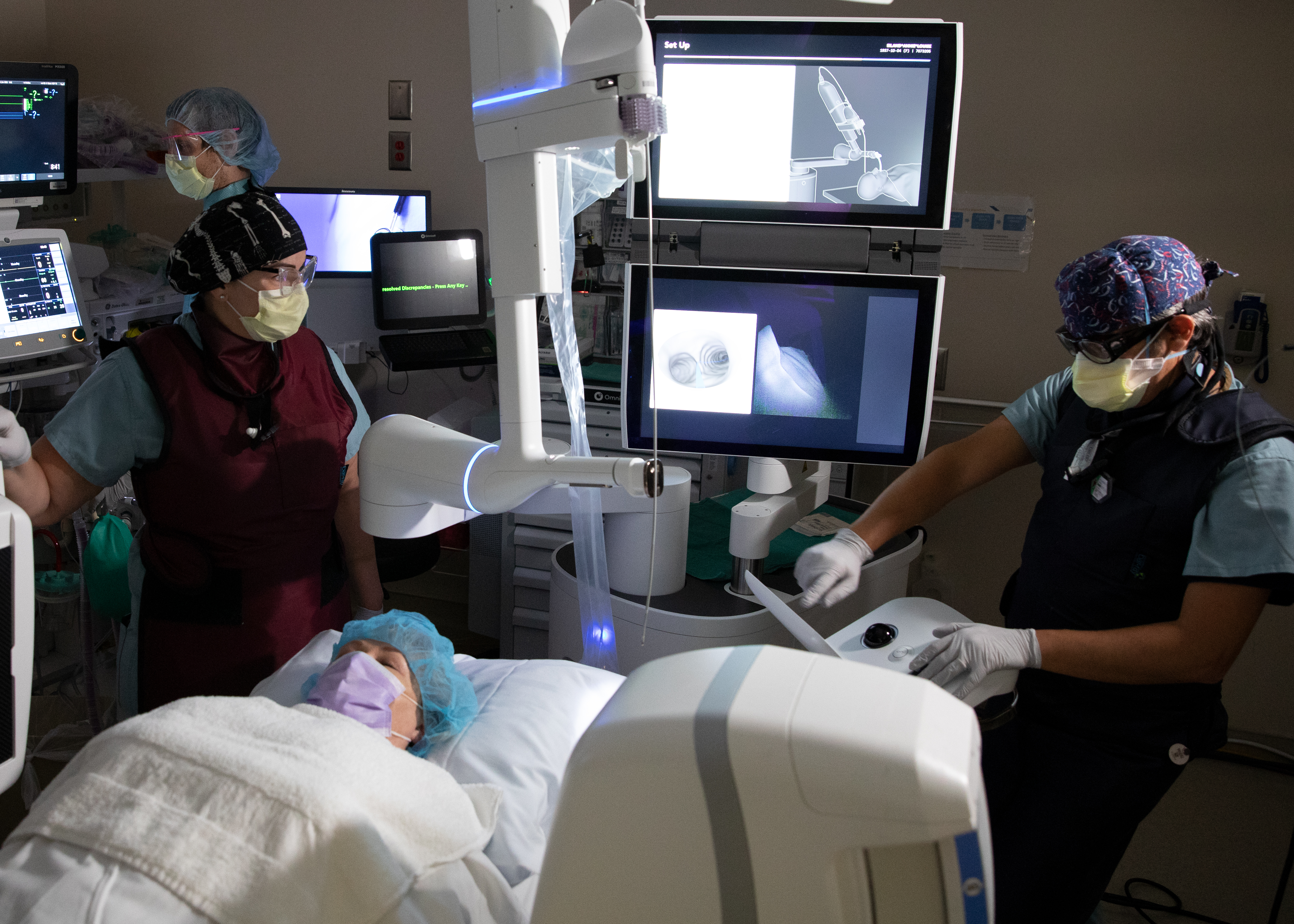
The mobile C-arm imaging system, called Cios Spin and produced by Siemens Healthineers, allows doctors to pinpoint the exact location of a suspicious lung nodule. Doctors at the hospital use the imaging system in conjunction with the Ion bronchoscopy robot, made by Intuitive, which allows them to guide an ultra-thin and maneuverable catheter deep into the peripheral areas of the lung to biopsy suspect tissue.
“Differentiating benign nodules from malignant cancer is of great importance, since early lung cancer diagnosis and surgical resection — before malignancies metastasize to the regional chest lymph nodes or distant organs — can save someone’s life and allow a good long-term outcome,” says Alejandro Sardi, MD, assistant professor of medicine at Emory University School of Medicine and an interventional pulmonologist at Emory Saint Joseph's Hospital.
Doctors at Emory Saint Joseph's were the first in the state in 2021 to use the Ion robot, which uses a controller and a heads-up display to allow doctors to navigate the camera-equipped catheter along a pre-planned path through the lung airways. With its 3.5-milimeter size and ability to articulate 180 degrees in any direction, the Ion is capable of reaching all 18 segments of the lung. Once the targeted area is reached, a flexible needle is used to biopsy the suspect tissue.
Both cardiothoracic surgeons and interventional pulmonologists at Emory Saint Joseph's utilize the Ion, which can be deployed in either the hybrid operating room (a surgical suite that combines the capabilities of a cardiac surgery room and catheterization room) or diagnostic suites. The new Cios Spin adds even more precision when using the Ion robot in the diagnostic suite.
“Having real-time, intra-procedural CT-like images along with robotic bronchoscopy gives us a much higher likelihood of achieving a diagnosis though this minimally-invasive procedure,” Sardi says.“Performing lung nodule biopsies though the airways, is not only safer, but it also has other advantages over the historical percutaneous CT guided biopsies, such as avoiding the possibility of lung collapse.”
The system also allows doctors to also determine a stage of the disease when tissue is discovered to be cancerous, ruling in or out the possibility of regional lymph node metastasis during the same intervention.
“Providing cancer diagnosis and lymph node staging in a single procedure is very convenient, as it can save time for the patient and avoid other unnecessary procedures,” Sardi says.“We are excited to be the first in the state to offer such minimally-invasive diagnostics in a single procedure.”