A collaborative effort between investigators at the Yerkes National Primate Research Center, Emory University School of Medicine and Georgia Institute of Technology has led to the development of a non-invasive method to image simian immunodeficiency virus (SIV) replication in real-time, in vivo.
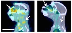
This approach, reported Monday in Nature Methods, is based on immune positron-emission tomography/computed tomography (PET/CT) and allows for the capture of viral dynamics of SIV, the animal model of human HIV infection. This novel approach has broad application to the study of immunodeficiency virus pathogenesis and drug and vaccine development, as well as to the potential use with human patients to identify viral reservoirs potentially leading to a cure of HIV/AIDS.
Francois Villinger, DVM/PhD, a researcher in the Yerkes Research Center’s Microbiology and Immunology division, and Philip Santangelo, PhD, a researcher at Emory University and in the Wallace H. Coulter Department of Biomedical Engineering at Georgia Institute of Technology, spearheaded the study with their respective teams and collaborators at the Emory School of Medicine. Using the nonhuman primate model of human HIV infection, their approach uncovered novel, previously unappreciated sites of viral replication, such as in nasal-tissue and the reproductive organs.
In addition, the methodology was able to capture the previously unappreciated wide variation in viral replication levels within select organs, including sections of the gastrointestinal tract, whether or not the subject was taking antiretroviral therapy (ART). Finally, the methodology allows for repeat analysis of the viral dynamics, for example during acute infection, during ART and upon cessation of ART.
"Before we can hope to eliminate reservoirs of infected HIV cells, we must first identify tissue sites that can possibly serve as these viral reservoirs," says Villinger. "With this new imaging method, we believe we can now do this more effectively in our animal models and that this strategy can translate these non-invasive techniques into investigating the eradication of HIV infection and targeting of virus reservoirs in humans," he continues.
"Use of the technique could lead to a better understanding of viral dynamics in the body, which could help target new generations of therapeutics and diagnostics," adds Santangelo. "This could help us find the regions where the virus is replicating and allow us to focus molecular diagnostics on the areas that are really important," he says.
The imaging tools developed in this study could also have potential applications with other pathogens and disease states, helping researchers understand where the infectious agents reside and how they respond to therapy. The researchers are developing new imaging probes for applying the same technology in humans in the near future.
Yerkes National Primate Research Center
Established in 1930, the Yerkes National Primate Research Center paved the way for what has become the National Institutes of Health-funded National Primate Research Center (NPRC) program. For more than eight decades, the Yerkes Research Center has been dedicated to conducting essential basic science and translational research to advance scientific understanding and to improve human health and well-being. Today, the Yerkes Research Center is one of only eight NPRCs. The center provides leadership, training and resources to foster scientific creativity, collaboration and discoveries, and research at the center is grounded in scientific integrity, expert knowledge, respect for colleagues, an open exchange of ideas and compassionate, quality animal care.
In the fields of microbiology and immunology, infectious diseases, pharmacology and drug discovery, transplantation, neurologic and psychiatric diseases, as well as behavioral, cognitive and developmental neuroscience, Yerkes scientists use innovative experimental models and cutting-edge technologies to explore and test transformative concepts aimed at: preventing and treating viral diseases such as AIDS; designing novel vaccines for infectious diseases such as malaria and tuberculosis; enhancing the potential of organ transplantation and regenerative medicine; discovering new drugs and drug classes through high-throughput screening; defining the basic neurobiology and genetics of social behavior and developing new therapies for disorders such as autism and drug addiction; understanding the biology of neurodegenerative diseases, such as Alzheimer’s and Parkinson’s diseases; and advancing knowledge about the evolutionary links between biology and behavior.
Coulter Department of Biomedical Engineering
The Wallace H. Coulter Department of Biomedical Engineering at Georgia Tech and Emory University is the largest biomedical engineering department in the United States, with 38 faculty, 1,400 undergraduate students and 200 graduate students. Operated in a partnership between a leading public engineering school and a highly respected private medical school, the Coulter Department’s graduate program is ranked second in the nation by U.S. News & World Report.