Bring in the Laser
Precise ablation technique shows progress in treating intractable epilepsy

Ja’Lisa Thomas was preparing dinner when the seizure struck. She fell forward, unconscious, over the hot electric stove. Only the quick reaction of her father, in pulling her to safety, prevented a catastrophe.
“I ended up burning my shirt and my arms, but luckily it was nothing too serious,” she remembers. “It could’ve been so much worse—the house could have gone up in flames.”
Thomas had her first seizure eight years ago, while living in Delaware and eight months pregnant with her daughter.
“I was sitting at my computer searching for information on the internet when my mother noticed that I had zoned out and was staring at the ceiling,” Thomas recalls. “She said I began to convulse and gasp for air. She called for my father to help get me to the floor and on my side.”

Ja’Lisa Thomas’ epileptic seizures are finally under control after a surgery at Emory Brain Health Center that involved precise laser ablation (elimination) of small areas of her brain where the seizures were originating.
Neither she nor her doctor knew the cause of this or subsequent seizures. They thought there might be a connection to her pregnancy. But the seizures persisted after her child was born, slowly increasing in frequency.
In 2013 Thomas and her family moved to the east metro-Atlanta area, and she sought answers at Emory, where she received a diagnosis of epilepsy.
Epilepsy is characterized by seizures that may result in a loss of consciousness, involuntary jerking movements of the arms and legs, temporary confusion, or a staring spell. The episodes can last anywhere from a few seconds to several minutes.
The symptoms appear to be a loss of control or function, suggesting disorganized cellular signaling in a particular part of the brain. In fact, it is just the opposite: the neurons are too aligned and are all firing at the same time and overloading the nerve pathways.
“Think about it this way,” says neurosurgeon Robert Gross, MBNA Bowman Chair in Neurosurgery and professor in Emory’s Department of Neurosurgery. “If you go into a theater and some people are whispering in a low voice, there’s no problem. If everyone whispers the same thing at the same time, the sound would be deafening.”
There is no medical cure for epilepsy, which affects an estimated three million people in the U.S., although sometimes it goes away on its own.
By the time Thomas was diagnosed, her seizures were occurring several times a month, sometimes back to back, even while she slept. One medication after another was tried, but nothing worked. She could no longer drive, and she couldn’t work because her condition forced her to take too many days off.
The worst part, Thomas says, was the effect her medical problems were having on her daughter, a toddler at the time.
“She was getting worried about me to the point where she felt she had to take care of me,” Thomas recalls, “and that broke my heart.”
Something had to change.
Having exhausted the standard options, Emory neurologists referred her to Gross, who specializes in “intractable” cases—the one-third or so of epilepsy patients for whom medication doesn’t work, leaving surgery as the only course of action.
The pre-surgical process, which can take between six months to a year or more, begins with a combination of tests and clinical considerations to determine where the seizures are likely to be originating.
“We start with the description from the patient of his or her own seizures and then we bring them into the hospital and allow or even provoke them to have their typical seizures while we videotape them,” Gross says.
During this process, electroencephalography sensors are positioned on the patient’s scalp to reveal correlations between the onset of seizure behaviors and neural activity in specific parts of the brain.
“We also perform MRI scans to see if there are any structural abnormalities in the brain,” Gross says. “Sometimes we see a lesion of some sort, a scarring or even a tumor or some other developmental abnormality that helps us develop our hypothesis as to where the seizures are arising. Sometimes we see no abnormalities at all.”
Further tests look for metabolic changes in brain areas that show correlations, and “we use neuropsychological tests of patients’ strengths or weaknesses with respect to their cognitive, emotional, and other neurological functions that can also help give us more clues as to where the seizures might be originating.”
This data is put together to form testable hypotheses as to which areas of the brain are responsible for the seizures and whether surgery would help.
With the brain’s suspected problem areas mapped out on a computer in 3-D, tiny holes are drilled into the skull at those areas.

Ja’Lisa Thomas’ epileptic seizures are finally under control after a surgery at Emory Brain Health Center that involved precise laser ablation (elimination) of small areas of her brain where the seizures were originating.
Ja’Lisa Thomas’ epileptic seizures are finally under control after a surgery at Emory Brain Health Center that involved precise laser ablation (elimination) of small areas of her brain where the seizures were originating.
In the Room
In Thomas’ case, 15 openings were made, and electrodes were inserted into her temporal, frontal, and parietal lobes to monitor brain activity and map seizures to either confirm or disprove the physicians’ hypotheses.
This process takes weeks in the hospital under 24/7 videotaped monitoring. During the monitoring period, electrical stimulation mapping is also performed, in which a small amount of electrical current is passed through the electrodes to “map out” the function of the brain near those electrodes.
Sometimes, with the patients’ consent, doctors may use this opportunity to conduct memory tests connected to other research projects (see sidebar, below main story).
At this point, all the clinical information is presented and reviewed at a comprehensive epilepsy conference attended by the neurologists, neurosurgeons, and neuropsychologists.
In Thomas’ case, two small areas in the frontal and temporal lobe were identified as the areas where her seizures were originating.
Then came the intervention: While Thomas was awake and being monitored for effects, those spots were heated and destroyed, using radio frequency waves—a laser ablation technique that can be sufficient to stop some patients’ seizures. In Thomas’ case, over the next twenty-four hours she continued to have seizures. More needed to be done.
So, a few days later, she was placed in the MRI scanner where the electrodes were withdrawn and replaced with a catheter containing a strand of glass optical fiber through which intense laser light is shined. This allowed for precise ablation of the epileptic brain tissue. Thomas’ procedure lasted about ten hours, but she was able to go home in a few days.
Until the advent of laser ablation about seven years ago, the go-to surgical procedure was an open brain resection, such as frontal or temporal lobectomy, which involved opening a section of skull and physically removing suspect brain tissue.

Neurosurgeon Robert Gross, MBNA Bowman Chair in Neurosurgery and professor in Emory’s Department of Neurosurgery, specializes in “intractable cases” of epilepsy, in which medications don't work.
“Ablation via precisely positioned lasers has become a more and more prevalent technique,” says Gross, who was among the first neurosurgeons in the world to adopt the procedure and remains a leading proponent for its use. “It allows us to accomplish the same end but with a minimally invasive and minimally destructive approach.”
As with any kind of brain surgery, laser ablation carries a certain level of risk, but the risk has to be weighed against the reward, notes Gross, whose surgical success rate hovers around 65 percent.
“In my group, we have demonstrated that in certain scenarios, there’s a decreased risk profile with ablation as compared to an open surgical treatment. And laser ablation is much more tolerable and comfortable for the patient, and that helps to increase the willingness of patients to undergo what is, in the end, life-altering surgery, and for neurologists to refer them for it.”
Thomas’ surgery was performed in May 2018.

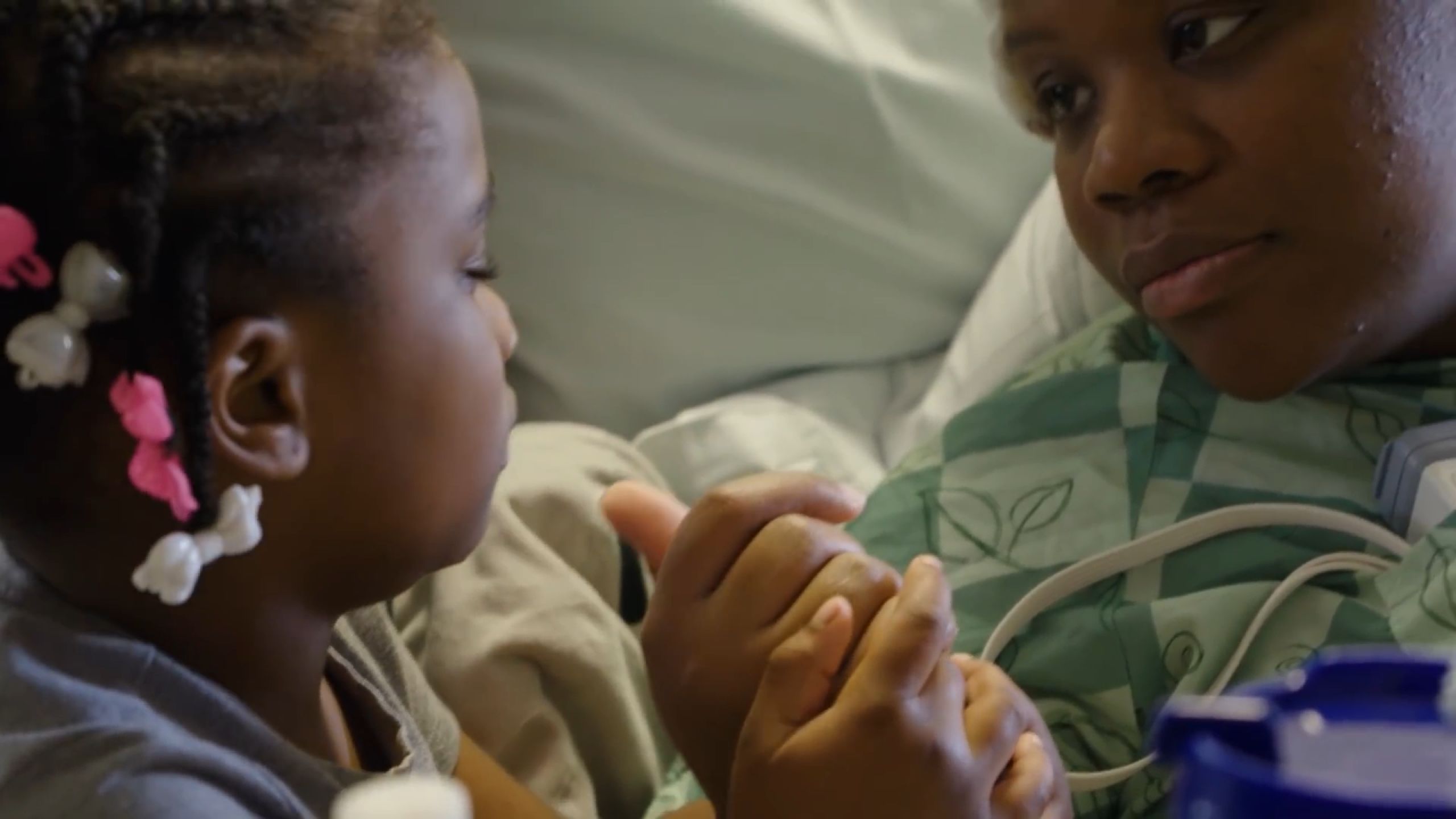
JaLisa Thomas, with her two daughters, has been able to resume normal daily activities after successful laser ablation surgery stopped her seizures.
“My life has changed a lot since then,” she says. She is driving again and found a job as an administrative assistant. Best of all, she is seizure free.
“Things are mostly back to normal and I’m very happy about that,” she says. “It was depressing to have to depend on other people to help me all the time, but I’m doing a lot of things on my own now.”
Written by Gary Goettling. Designed by Peta Westmaas. Edited by Mary Loftus.

Neurosurgeon Robert Gross, MBNA Bowman Chair in Neurosurgery and professor in Emory’s Department of Neurosurgery, specializes in “intractable cases” of epilepsy, in which medications don't work.
Neurosurgeon Robert Gross, MBNA Bowman Chair in Neurosurgery and professor in Emory’s Department of Neurosurgery, specializes in “intractable cases” of epilepsy, in which medications don't work.

JaLisa Thomas, with her two daughters, has been able to resume normal daily activities after successful laser ablation surgery stopped her seizures.
JaLisa Thomas, with her two daughters, has been able to resume normal daily activities after successful laser ablation surgery stopped her seizures.
Research
A 'Memory Pacemaker’
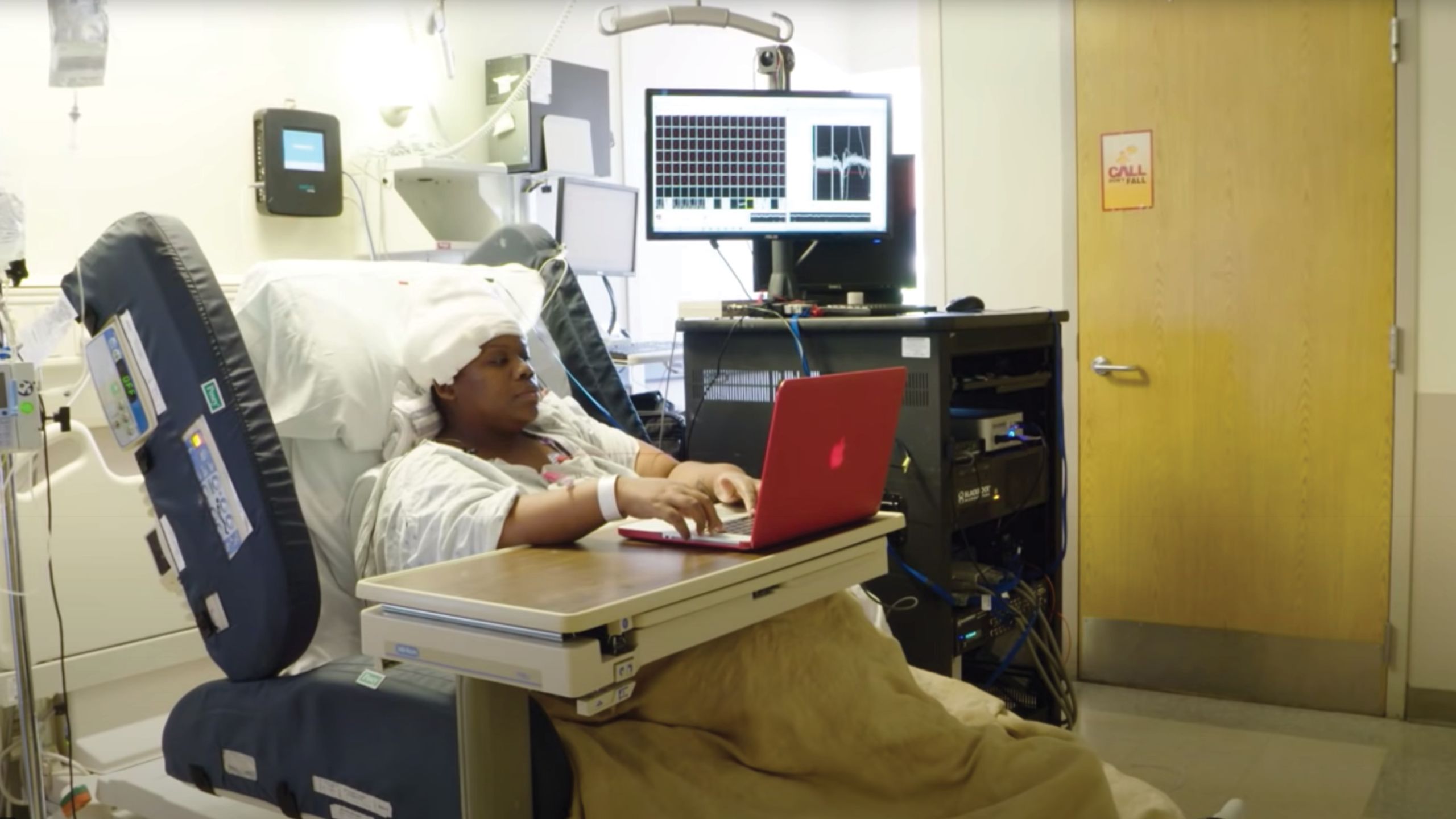
While in the hospital, Ja’Lisa Thomas volunteered to participate in the Restoring Active Memory program, a research project intended to study the electrophysiological signals associated with memory.
“We want to see if we can do something about improving those signals and memory functions,” says the project’s principal investigator, Robert Gross, MBNA Bowman Chair in Neurosurgery and professor in Emory’s Department of Neurosurgery.
“Epilepsy patients with electrodes going into their brain recording their brain activity, and who are also amenable to being stimulated electrically as part of routine epilepsy monitoring and care, are the perfect test subjects for learning about brain structures.”
Launched in 2013 under the auspices of DARPA (Defense Advanced Research Projects Agency), the program's goal is to develop an implantable, closed-loop neural interface capable of restoring normal memory function—in effect, a memory pacemaker.
Intended to help military personnel overcome the effects of brain injury or illness, the device could find wide civilian applications, assisting individuals with Alzheimer’s and other forms of dementia or deficits arising from traumatic brain injury.
Emory is one of eight institutions across the country involved in the collaborative effort.
Thomas participated in a memory test where she memorized lists of words and then, after a distraction, was asked to recall as many of the words as she could. Throughout the process, neural activity in specific areas of her brain known to play a role in memory was monitored and recorded.
The test was repeated several times with different words. Sometimes memorization was conducted while the memory-related areas were stimulated; other times, it was conducted without stimulation.
The idea behind the research is to determine the conditions under which targeted electrical stimulation of specific brain cells improves memory. The DARPA program memory tests dovetail with other research conducted by Gross and his team on the potential benefits of deep-brain stimulation.
In a study published in 2018, principal investigator and neurosurgeon Jon Willie, assistant professor in the Department of Neurosurgery, working with Gross and others, shared results showing that stimulation of the brain's amygdala can improve memory function.
Gross also has played an integral role in teams that have pioneered the development and successful implantation of devices that deliver electrical stimulation to precise targets in the brains of epilepsy patients.
Analogous to a cardiac pacemaker, these devices reduce and in some cases abolish seizures in individuals who do not respond to standard epilepsy medication and who are not candidates for destructive surgery such as brain resection or laser ablation.