The National Institute on Aging has awarded Emory researchers a five-year, $3.8 million grant to investigate advanced brain imaging techniques for Alzheimer’s disease.
Deqiang Qiu, PhD, and colleagues will investigate how problems in blood flow, known as cerebrovascular impairment, contribute to the early phases of Alzheimer’s. The goal of the project is to study how Alzheimer’s pathology interacts with cerebrovascular impairment in the early phases of the disease, as well as the relative contributions of cerebrovascular and Alzheimer’s pathologies to cognitive impairment.
“Alzheimer’s disease is a devastating condition that affects an ever-increasing number of patients and families,” says Qiu, who is associate professor of radiology and imaging sciences at Emory and associate professor in the Wallace H. Coulter Department of Biomedical Engineering at Georgia Tech and Emory. “I’m extremely excited and honored to start this project to better understand the role of cerebrovascular impairment, an important but under-studied factor in the Alzheimer’s disease process.”
Qiu is also director of the Department of Radiology’s Computational Neuroimaging and Neuroscience Laboratory and program director for magnetic resonance imaging (MRI) at Emory’s Center for Systems Imaging.
The project will include collaborations with Emory Goizueta Alzheimer’s Disease Research Center and the Emory Healthy Brain Study. Co-investigators include James Lah, MD, PhD, and Allan Levey, MD, PhD, in the Department of Neurology; Jason Allen, MD, PhD, and Jonathon Nye, PhD, in the Department of Radiology and Imaging Sciences and the Center for Systems Imaging; and Benjamin Risk, PhD, at Rollins School of Public Health.
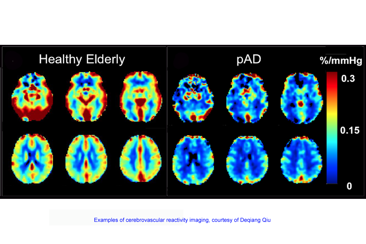
In the planned study, investigators will use a new MRI technique developed by Qiu’s lab that simultaneously measures cerebral blood flow and blood oxygenation levels. The technique involves temporarily exposing someone to air with normal levels of oxygen and slightly elevated levels of carbon dioxide.
“This will increase the blood flow and oxygenation level in a normal brain, termed cerebrovascular reactivity or CVR,” Qiu says. “However, CVR is impaired in aging and more severely impaired in Alzheimer’s disease.”
The MRI-based approach will be combined with positron emission tomography (PET) to image the deposition of amyloid plaques in the brain, a characteristic pathology seen in Alzheimer’s that also can appear in healthy individuals.
The study will include healthy younger and elderly participants, people who are asymptomatic but display amyloid plaques via PET imaging, as well as people who are considered to have a prodromal stage of Alzheimer’s because they have mild cognitive impairment and signs of Alzheimer’s pathology. Qiu says the project could eventually lead to the development of effective multi-component therapeutic interventions that target both AD and vascular pathologies.